Vertebral Column and Sacro Iliac Joint
Anotomy Lumbar Vertebral Column and Sacro-Iliacal Joint
An overview of the anatomy, in this case, the anatomy of the lumbar and sacral column, is in my view necessary to create a common starting point
The Vertebral Column, a stabilized axis
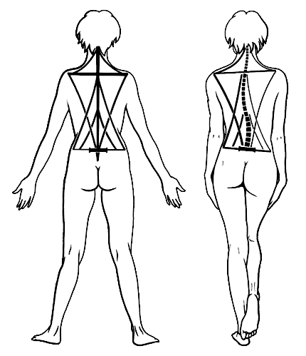
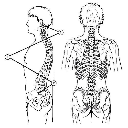
The spine can be seen as the mast of a ship, with lines at the mast to ensure its rigidity and flexibility (plasticity). This mast is resting on the pelvis, extends to the head and at the level of the shoulders there is a main-yard set transversely, that is the shoulder girdle. At all levels there are ligaments and muscle tightening tensioners arranged as stays. Linking the mast to its attachment site, the pelvis. And at the level of the shoulder connecting the shoulder girdle to the vertebral column and the horizontal stay. In the upright posture, the active lines (musculature) and passive lines (ligaments) are in balance.
If the symmetry is lost, the pelvis tilts to one side and a sequence of convexity and concavity in the spinal column will occur, supported by muscles and ligaments, and the balance is maintained (extrapyramidal system). The flexibility of the spinal column is due to the stacked components, which are connected by (smaller) lines (muscles and ligaments). As a result, the spinal column, even when changing of shape, will stay rigid.
The Vertebral Column, an Axis and a Protector
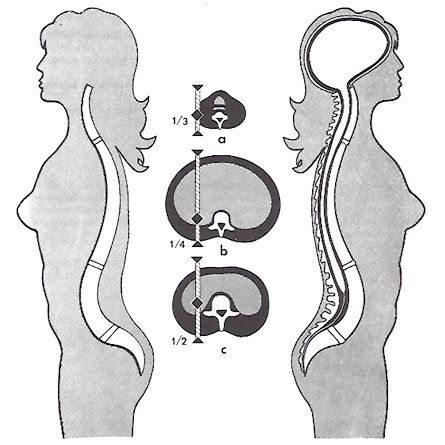
Although the position of the spine in the body is as a central axis, but it differs for each level. In the cervical spine, the spine should be as close as possible to the center of gravity of the head (⅓). In the thoracic spine, the spine must accommodate the internal thoracic organs (¼). The lumbar spine carries more weight, and will consequently also be as close as possible to the center of gravity (½).
An important function, in addition to the already mentioned parameters related to the biomechanics, is the protection that it should provide to the spinal cord. From the foramen magnum to the sacral canal. The structures used (and solutions) to ensure flexibility has made a concession in respect of the protection. Great flexibility gives less protection to the neurogenic structures. On the other hand provides a high degree of protection, a high rigidity.
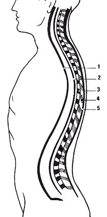
The Vertebral Column, Normal Curvatures
Normal concavities and convexities of the spinal column
The Vertebral Column, Structure of a Typical Vertebra
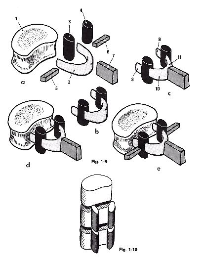
The building blocks of a vertebra. The basic form applies to all vertebrae, but with different components may have undergone great changes. An important aspect is that, by stacking these blocks together, there are three pillars been developed. One pillar at the front side is formed by the vertebrae corpora lying one above the other.
2 smaller pillars created by the processus aticulares lying one above the other.
The vertebral canal arises by the boundary at the front side by the vertebral body, and on the back side of the vertebral arch. By stacking the vertebrae there arises the vertebral canal, which is alternately bounded osteogenic or ligamentous.
The Vertebral Column, the Curvatures and Pressure Resistance
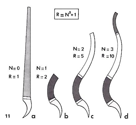
The resistance to compressive forces in the axial direction is increased by the curvature in the spine. The resistance equals to the square of the number of curves plus one.
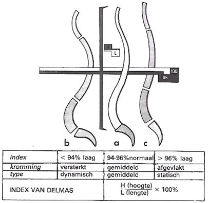
The index of Delmas indicates the strength of the curvatures of the spine. The ratio between the height and the length of the spine (from S1 to cover plate atlas).
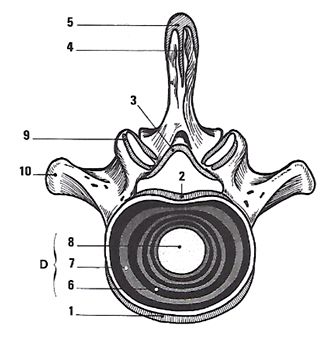
The Vertebral Column, the Linkage between Vertebrae
- 1 lig longitudinale anterius (skull base --> sacrum)
- 2 lig longitudinale posterius (pars basillaris --> canalis sacralis)
- 3 lig flavum (thick and tough; associated with each vertebra)
- 4 lig interspinale (connected to the lig supraspinale)
- 5 lig supraspinale (attached to the ligamentum supraspinal)
- 6 discus intervertebralis
- 7 discus intervertebralis
- 8 discus intervertebralis
- 9 lig anterius and lig posterius
- 10 lig intertransversarium
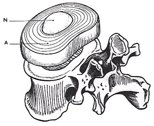
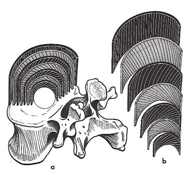
The Vertebral Column, the Structure of the Intervertebral Disc
Central section:
N is the nucleus, gelatinous, embryologically descendent of the chorda dorsalis, is made up of 80% water and is very hydrophilic due to Hyaluronan.
Peripheral part:
A is the annulus fibrosus, a fibrous ring made up of concentric lamellae.
The nucleus is enclosed by an inextensible space formed by the closure plates of the vertebrae and the annulus fibrosus.
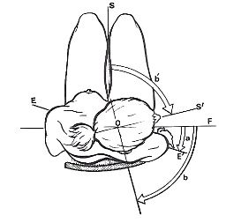
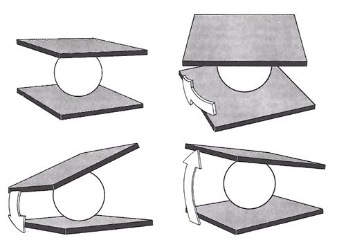
The Vertebral Column, the Nucleus Pulposus as a Swivel
There are 3 types of motion as possible at this "ball joint";
A: bending: in the sagittal plane: flexion vs. extension. In the frontal plane: lateral flexion
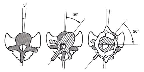
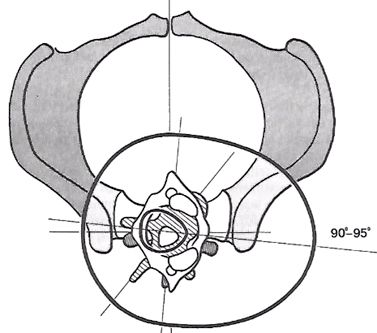
B: Rotation:
C: sliding
This results in six degrees of freedom:
Flexion / Extension
Lateral flexion left and right,
sliding in sagittal plane
sliding in frontal plane
rotation to the right
rotate to the left
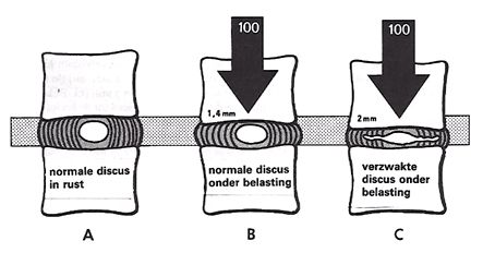
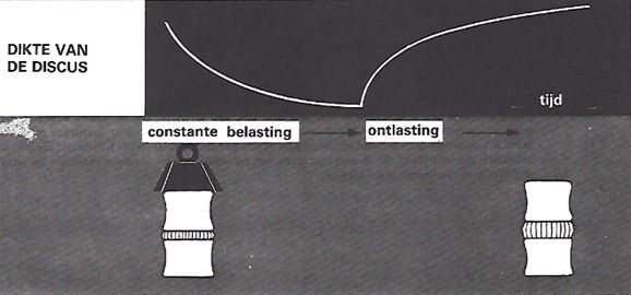
The Vertebral Column, Water Imbibition by the Nucleus
When loading the spinal column, the water in the nucleus moves by pores in the cover plate into the cancellous bone. In resting position the water returns back through the hydrophilic nature of the nucleus.
The pressure forces on the disc are increasing towards the sacrum. More weight should be worn.
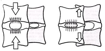
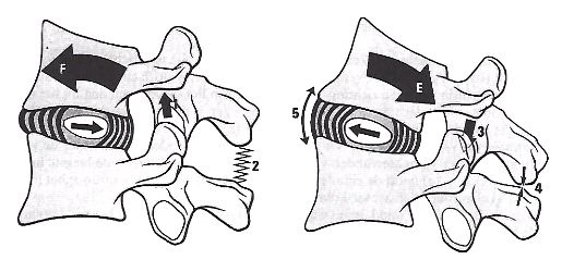
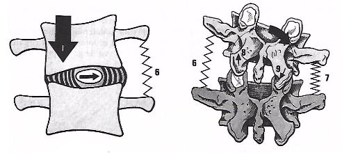
The Vertebral Column, Disc Behaviour during simple Movements
One can clearly see the self-stabilizing capability of the intervertebral disc. The nucleus moves in an opposite direction and increasing tension on the annulus and this tends to restore the upper vertebra to its original position.
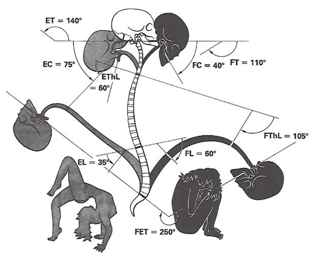
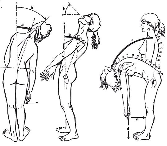
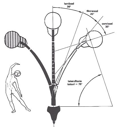
The Vertebral Column, Range of Movements